Hysteroscopy and treatment of the uterine factor
Hysteroscopy is a minimally invasive technique in which a video camera is introduced into the uterine cavity, and the cavity is distended with saline solution to allow adequate visualization. It serves both as a diagnostic technique for intrauterine pathology and as a treatment. With microscissors, micro-forceps, or a diathermy loop, polyps and fibroids can be removed, biopsies can be taken, the uterine cavity can be enlarged, and more.
In addition to resolving intrauterine pathology that can cause pain and bleeding, hysteroscopy makes it possible to diagnose and treat uterine factors that can contribute to infertility or increase the risk of miscarriage. Some of these factors include:
- Uterine anomalies: Congenital or acquired abnormalities in the structure of the uterus can interfere with embryo implantation or hinder the proper development of the pregnancy. Examples of uterine anomalies are the septate uterus, the bicornuate uterus, the didelphic uterus, or the presence of uterine polyps.
- Uterine fibroids: Fibroids are benign tumors that develop in the muscular wall of the uterus. Depending on their size and location, they can interfere with embryo implantation or cause complications during pregnancy, such as miscarriage or preterm birth.
- Uterine synechiae: Also known as uterine adhesions or Asherman syndrome, this refers to the formation of scar tissue inside the uterus. These adhesions can result from previous uterine surgery, infections, or trauma, and can interfere with implantation and embryo development.
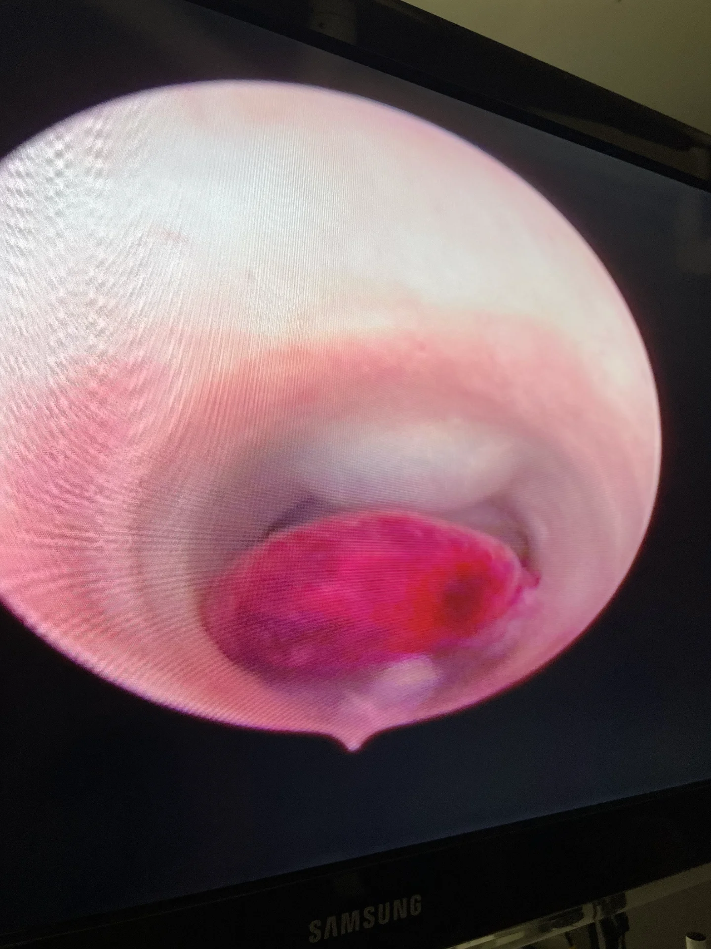
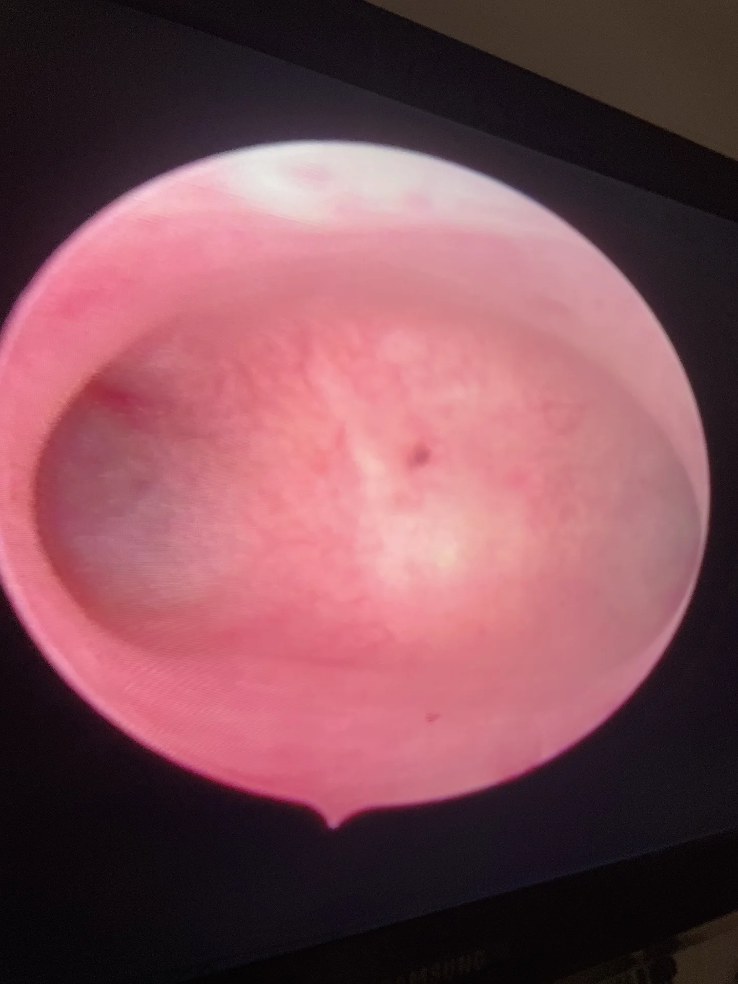
- Adenomyosis, which is endometriosis of the uterus; that is, when endometrial tissue infiltrates the muscular layer of the uterus, or myometrium.
- Uterine infections: Uterine infections, such as pelvic inflammatory disease (PID), can damage the fallopian tubes and uterine tissue, making it harder for the fertilized egg to be transported properly or increasing the risk of miscarriage. Chronic endometritis is usually of infectious origin and clearly reduces the implantation rate. Hysteroscopy facilitates the diagnosis of endometritis, first because it detects highly suggestive signs such as micropolyps, diffuse or focal edema, and hyperemia, especially when it has a strawberry pattern. The second advantage of hysteroscopy in diagnosing chronic endometritis is that it allows a targeted biopsy to be taken to confirm the diagnosis.
I perform hysteroscopies at C/ Villa de Marín, CNN 1328021423, and at Hospital San Francisco de Asís, on Calle Joaquín Costa, CCN: 132800005