Cervical pathology and colposcopy
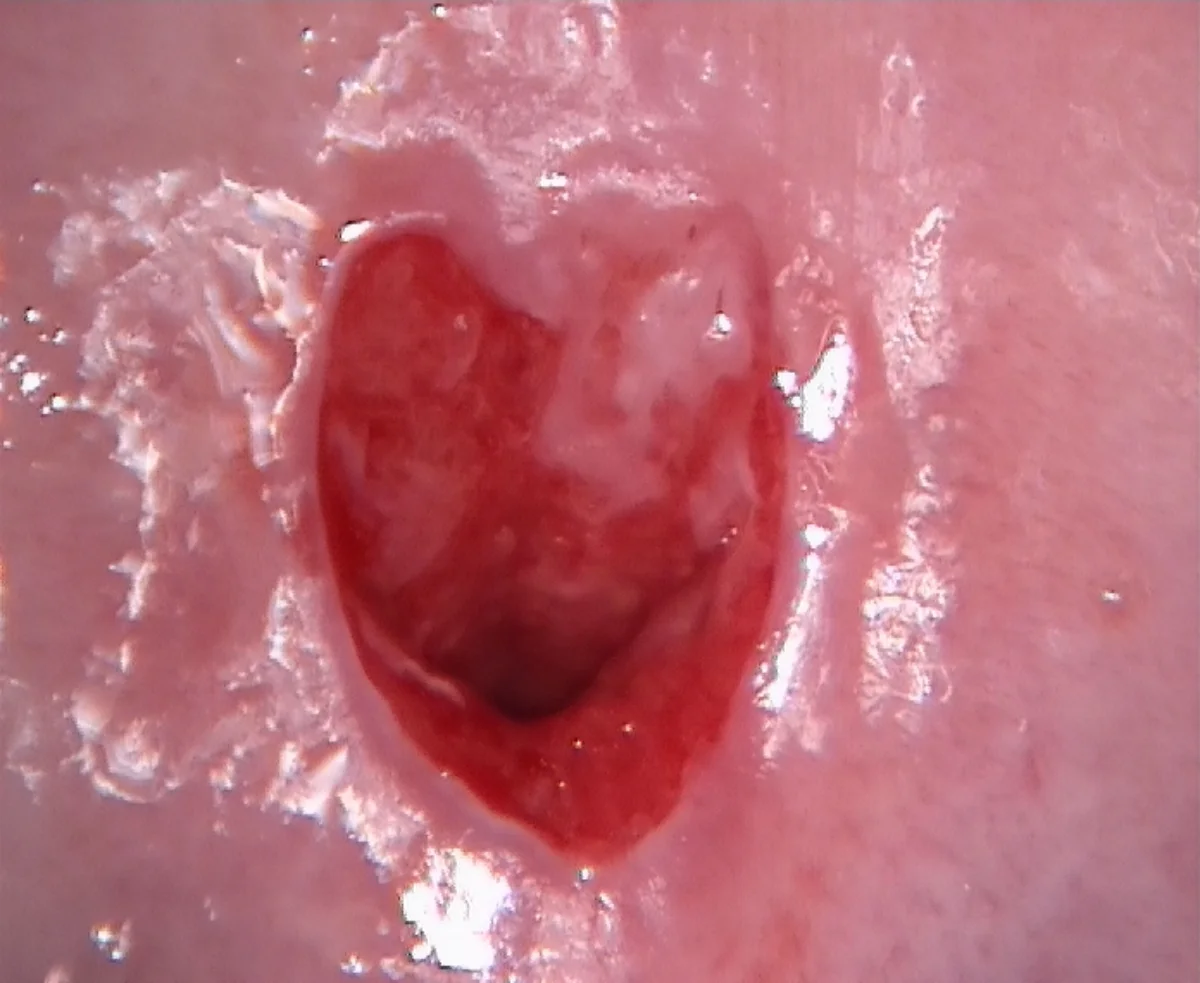
Cervical pathology and colposcopy. According to the World Health Organization (WHO), cervical cancer is the fourth most common cancer in women and causes more than 300,000 deaths a year. The main cause is persistent infection with high-risk strains of the human papillomavirus (HPV). Vaccination does not protect against all high-risk HPV strains, so screening remains essential for early diagnosis and for treating premalignant lesions and cancer at stages when they can still be cured.
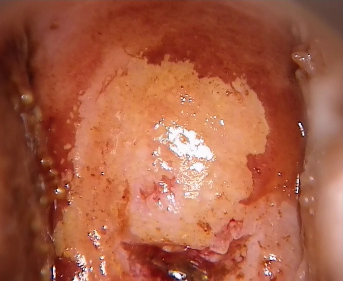
Colposcopy consists of viewing the cervix with a magnifying lens and applying acetic acid and Lugol’s iodine. When there are premalignant lesions on the cervix, characteristic images are seen through the lens, and the tissue stains in a particular way with acetic acid and Lugol’s iodine. Colposcopy is the “gold standard” technique for the early diagnosis of cervical cancer. It is especially important when the woman has a high-risk papillomavirus infection or when lesions are detected on cytology.
Although, according to protocols, it is not always essential, it is a harmless technique, and I include a colposcopy for all our patients at their annual check-up if they wish. The most important suspicious signs of a high-grade lesion on colposcopy are intense, raised acetowhite epithelium (ridge sign), atypical mosaic or punctation (coarse and irregular), irregular and fragile vascularization, absence of Lugol uptake, especially at the squamocolumnar junction, dense acetowhite on columnar epithelium, and the peel-off sign (tissue that detaches easily). If any of these signs is detected, it is essential to take a targeted biopsy for subsequent pathological study.