High-resolution and 3D ultrasound
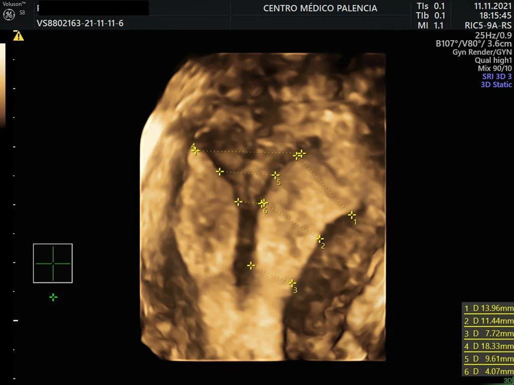
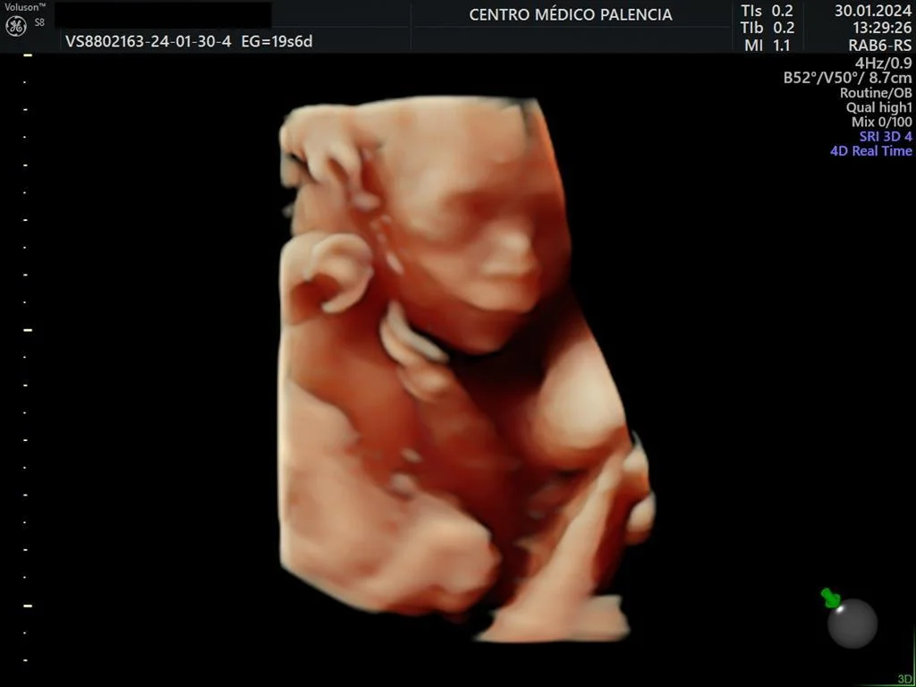
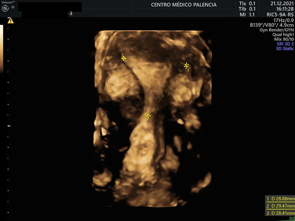
High-resolution and 3D ultrasound. Study of adenomyosis. Traditionally, 3D ultrasound has been used in prenatal diagnosis to assess fetal malformations. However, this is not the only field in which it offers great advantages. In gynecology, and especially in fertility, high-resolution ultrasound and three-dimensional reconstruction of the uterine cavity are particularly useful for diagnosing malformations of the uterine cavity, deep endometriosis, and adenomyosis.
Endometriosis is a condition in which the tissue lining the uterus, the endometrium, grows outside the uterus, usually on the ovaries, the fallopian tubes, or surrounding tissue. Endometriosis can affect fertility by blocking the fallopian tubes, causing inflammation and scarring, or affecting egg quality.
Adenomyosis is a disorder in which the endometrial glands invade the underlying myometrium. Adenomyosis usually causes abdominal pain and changes in the bleeding pattern, but it is sometimes asymptomatic. Diagnosis of adenomyosis with low-resolution ultrasound machines can go unnoticed. The direct ultrasound criteria are intramyometrial cysts, hyperechoic islands, and subendometrial buds.
The indirect criteria are a globular uterus, myometrial asymmetry, fan-shaped shadowing, and an irregular junctional zone. Sensitivity for assessing the junctional zone increases especially with reconstruction of the uterine cavity in the coronal plane, which 3D ultrasound makes possible, with a resolution similar to that of pelvic magnetic resonance imaging. Doppler assessment of uterine vascularization, which high-resolution machines allow, also helps in the differential diagnosis between myoma and adenomyosis.
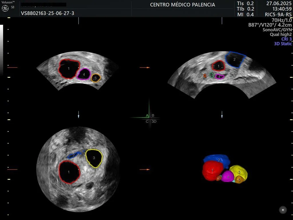
Adenomyosis reduces the chances of success in fertility treatment by 25-30%, so it is very important to take it into account in order to give a realistic prognosis. When monitoring ovulation stimulation, modern SonoAVC software allows automated counting and three-dimensional measurement of all follicles. Finally, the resolution of the ultrasound machine helps distinguish benign adnexal cysts from malignant ones. Ultrasound criteria of malignancy are irregular walls (>3 mm), thick septa, solid papillary projections, abundant internal vascularization detected by color Doppler, and a heterogeneous appearance with refractile areas.
High-resolution 3D ultrasound is available at every consultation held at Lagasca 122 (health authorization code CS2554)